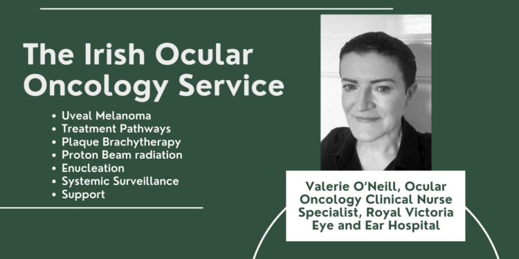
In 2010 a dedicated ocular oncology service was established in Ireland at The Royal Victoria Eye and Ear Hospital (RVEEH) in conjunction with St Luke’s hospital (SLH) in Rathgar. Prior to this all patients were referred to the United Kingdom for management of Uveal Melanoma (UM).

Written by Valerie O’Neill, Ocular Oncology Clinical Nurse Specialist, Royal Victoria Eye and Ear Hospital
The multidisciplinary team, delivering this service, is composed of an ocular oncology consultant, a radiation oncologist, a medical ophthalmologist, two residents, Physicists, we work closely with the pathologist and local medical oncologists, a Clinical Nurse Specialist (CNS), a Medical Social Worker (MSW), Eye Clinic Liaison Officer (ECLO) and our administration support.
Uveal Melanoma
Uveal melanoma is the most common primary intraocular malignancy in adults, albeit still a rare form of cancer. Melanoma arises from the pigmented cells (melanocytes) of the uvea. The uvea is divided into three parts termed the iris, ciliary body, and choroid. The uvea is comprised of blood vessels and melanocytes. A malignant melanoma of the uvea originates from these melanocytes, in the choroid, ciliary body or iris.
Uveal melanoma may not cause any signs or symptoms and is commonly found incidentally, detected during routine examination. Symptoms may include decreased or blurry vision.
There Is no known cause of UM. However, it is more common in those who are fair skinned and have light eye colour.
Treatment Pathways
Nationwide referrals are received by this dedicated oncology clinic, patients are referred with a suspicious naevus or ocular lesion for specialist investigation either via optician’s, another ophthalmologist, or through our own casualty department. A small minority come from the diabetic screening national service.
A diagnosis of uveal melanoma is made based on clinical findings from a dilated fundus examination and multimodal imaging, which consists of colour fundus photography optical coherence topography and B-Scan ultrasound. Treatment pathway decisions are based on tumour thickness and tumour location. These pathways include Brachytherapy for tumours that are less than 10mm, Proton beam radiation for tumours that are small and are close to the optic nerve, Enucleation removal of the globe and its contents for larger tumours, or specific laser treatment for smaller tumours known as transpupillary laser therapy (TTT) or Photodynamic therapy (PDT).
Treatment of uveal melanoma has evolved over the last four decades with eye conserving treatment options such as proton beam radiation and brachytherapy. Despite the therapeutic shift enucleation is still undertaken for larger tumours not amenable to radiation treatment.
The most commonly used care pathways are plaque brachytherapy, which is conducted in SLH, Enucleation which is performed in RVEEH and Proton beam radiation.
Plaque Brachytherapy
This treatment requires hospital admission for up to a week and includes two surgical procedures with general anaesthesia, insertion, and subsequent removal of the plaque once the radiation dose has been delivered.
This is a globe saving treatment option. Plaque Brachytherapy is used for uveal melanomas up to 10mm in thickness. In general ruthenium plaques Ru-106 are used to treat tumours up to 5mm in thickness and iodine plaques I-125 are used to treat tumours between 5-10mm. Plaque brachytherapy is described as a high intensity localised tumour control.
Proton Beam radiation
This treatment pathway is used for Uveal Melanomas located in the peripapillary or juxtapupillary region, these melanomas are close to the optic nerve. These patients are referred to St Pauls Eye hospital Liverpool and the Clatterbridge cancer care centre. This treatment is used to treat smaller tumours and is highly effective in achieving local control. All proton beam patients are treated in the UK, but all follow up care is completed in RVEEH.
Enucleation
This care pathway treats larger uveal melanomas measuring greater than 10mm in thickness. These larger tumours are not amenable to radiation.
Enucleation is removal of the globe and its contents and insertion of a volume replacing implant, leaving behind the muscles of the eye which support movement of the prosthesis once fitted. This procedure is performed in RVEEH. The patient stays overnight in RVEEH and is discharged with a pressure dressing in place for one week. They return to a nurse led clinic for removal of this dressing one week after surgery.
The patient is seen five weeks later to ensure the sutures are dissolved and they are ok to proceed with the prosthesis fitting. Development of the ocular oncology service over time has given us the scope to set up nurse led clinics which are specifically for the post-operative enucleation patient where these patients are seen in a dedicated clinic run by the CNS for all subsequent appointments.
Systemic Surveillance
Despite the availability of multiple treatment options, the survival for uveal melanoma patient has not changed for decades.
Approximately 50% of uveal melanoma patients will develop a metastatic disease within a 5–10year period. All treated patients within the service are referred to a medical oncologist locally for systemic surveillance. The liver is the most common site in the body affected by metastasis of an ocular melanoma and is often associated with poor prognosis.
Support
Patient support in the service includes a CNS who can be contacted directly, excellent support from our medical social work department who links with local cancer support services or psycho-oncology supports if needed. We also have direct links with the Irish cancer society and have run the cancer thrive and survive programme (CTS) on site with a view to running this again in the coming year.
The CTS programme focuses on problems common to cancer survivors and teaches coping strategies through action planning, feedback, behaviour modelling, problem solving and decisionmaking techniques. Family members, friends and caregivers are also welcomed to participate. This programme provides information and teaches practical skills on how to manage health care issues. CTS gives people the confidence and motivation they need to manage the challenges of living with a cancer diagnosis and meet people socially with similar needs and experiences.
Uveal melanoma and its treatment can influence the physical and psychological well-being of patients in a way that differs from other cancers. Factors to consider are visual impairment changes in appearance day to day function ocular discomfort and worry regarding disease recurrence. We are committed to excellence in care and to support the UM patient along the care pathway.
View the February Magazine here


